Understanding the Importance of a CT Scan for Lung Cancer Detection and Its Role in Modern Healthcare
In the realm of contemporary health and medical practices, the utilization of advanced diagnostic tools has revolutionized early detection and treatment planning for numerous diseases. Among these, the CT scan for lung cancer stands out as a pivotal technology that offers unparalleled accuracy, speed, and non-invasiveness. This comprehensive article explores the nuances of CT scans, their critical role in diagnosing lung cancer, and how such technologies are shaping the future of healthcare at leading centers like Hellophysio.sg.
What is a CT Scan and Why Is It Vital in Medical Diagnostics?
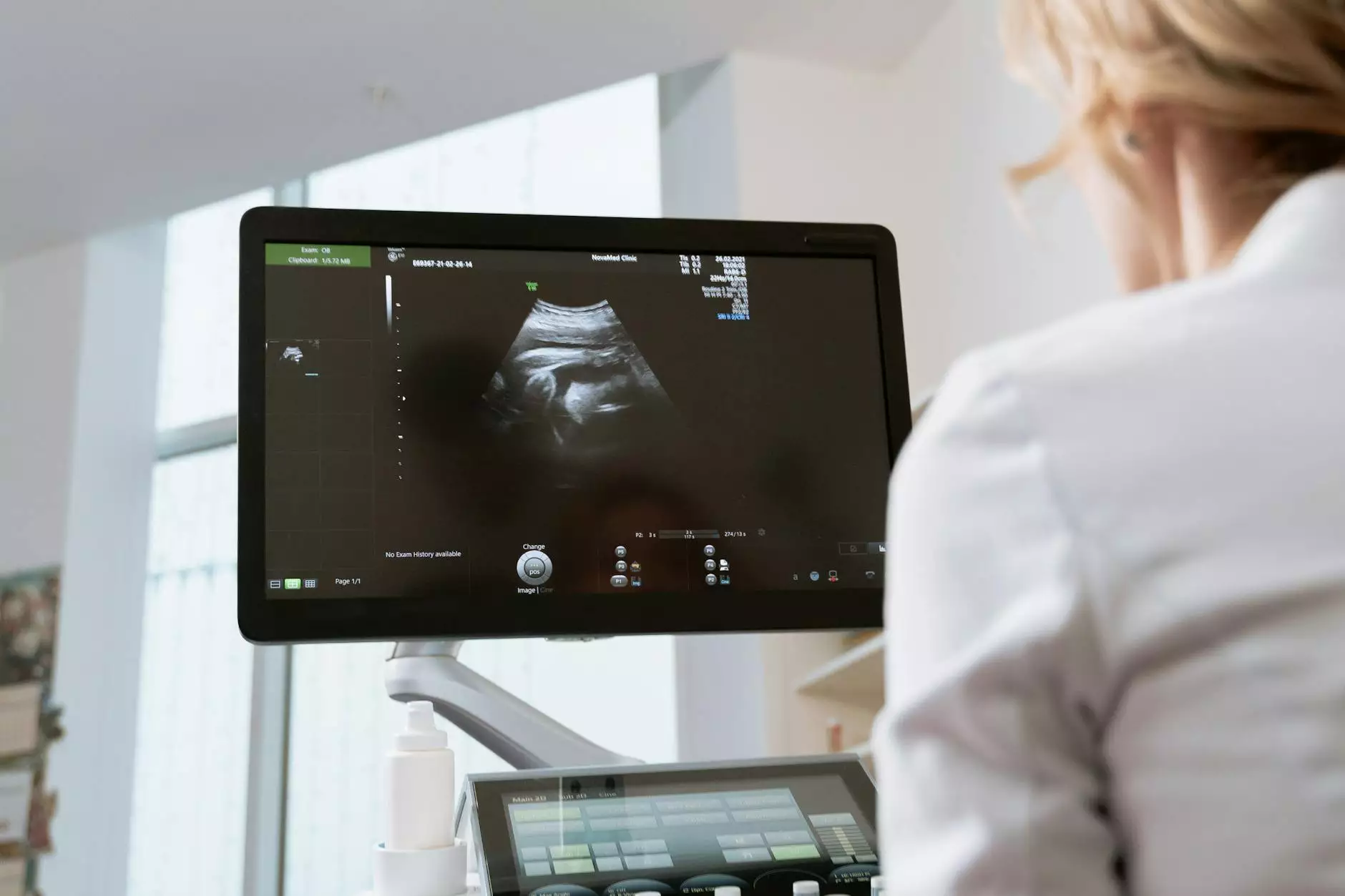
A CT scan, also known as computed tomography, is a sophisticated imaging technique that produces detailed cross-sectional images of the body. Using specialized X-ray technology and computer processing, a CT scan reveals the internal anatomy in stunning detail, highlighting abnormalities that may not be visible through conventional radiography.
The precision of a CT scan for lung cancer is particularly critical because early detection dramatically increases the chances of successful treatment. It helps physicians visualize small nodules or tumors within the lung tissues that might be missed on a traditional chest X-ray. The ability to see detailed 3D images assists in precise localization, staging, and even biopsy guidance for potential cancerous growths.
The Role of a CT Scan in Lung Cancer Screening and Diagnosis
Early Detection Enhances Treatment Outcomes
One of the foremost benefits of deploying a CT scan for lung cancer is its role in early diagnosis, particularly in high-risk populations such as heavy smokers or individuals with a history of certain lung diseases. Detecting lung tumors at an early stage significantly improves the prognosis and expands the options for effective treatments.
How Does a CT Scan Work for Detecting Lung Cancer?
The process involves the patient lying on a motorized table that moves through the CT scanner's circular opening. Through rapid rotating X-ray beams, the scanner captures numerous images from different angles. The images are then compiled by sophisticated software to form detailed cross-sectional views of the lungs.
- High-resolution imaging: Enables detection of small nodules as tiny as 1-2 millimeters.
- Volumetric data acquisition: Facilitates 3D reconstructions for precise localization.
- Enhanced contrast options: Allow differentiation between different tissue types, identifying suspicious masses.
Diagnostic Accuracy Compared to Other Imaging Modalities
Compared to traditional chest X-rays, CT scans for lung cancer demonstrate significantly higher sensitivity and specificity. While chest X-rays may miss small or centrally located tumors, CT imaging provides a comprehensive view, minimizing false negatives. The detailed visualization supports not only detection but also staging, which is essential for planning appropriate interventions.
Preparation and Procedure for a CT Scan of the Lungs
What to Expect Before the Scan
- Medical history review: Your healthcare provider will assess your health status and any allergies, especially to contrast materials if they are to be used.
- Fasting guidelines: Usually, fasting for a few hours before the scan is recommended if contrast dye is involved.
- Clothing and accessories: Wear comfortable clothing, avoiding jewelry or metallic items that could interfere with imaging.
During the Scan
The procedure is quick, typically lasting between 10 to 30 minutes. You will lie flat on the table while the scanner makes whirling movements. Patients are asked to hold their breath briefly to avoid motion artifacts, ensuring clear images.
Post-Procedure Care
If contrast dye was used, you may be advised to hydrate well to flush out the dye. There are generally minimal risks, but inform your provider if you experience any allergic reactions or discomfort.
Advantages of Using a CT Scan for Lung Cancer Diagnosis
- High sensitivity: Capable of detecting small lung nodules early on.
- Non-invasive: Does not require surgical intervention or biopsy initially.
- Speed and efficiency: Rapid results support timely decision-making.
- Guides further intervention: Aids in biopsy, surgery planning, and radiotherapy targeting.
- Monitoring disease progression: Follow-up scans evaluate treatment efficacy or detect recurrences.
Limitations and Risks of CT Scans
While CT scans are invaluable, they come with certain limitations. These include exposure to ionizing radiation, which although low, must be justified, especially for screening purposes. Additionally, false positives can occur, leading potentially to unnecessary biopsies or anxiety. Therefore, imaging should always be complemented with clinical assessments and other diagnostic tests.
The Significance of Accurate Interpretation in Lung Cancer Screening
Expert radiological interpretation of CT images is crucial. Radiologists trained in thoracic imaging identify suspicious lesions, assess their likelihood of malignancy, and determine the necessity for additional tests. Multiple factors such as nodule size, shape, edge characteristics, and growth rate influence diagnosis. The integration of artificial intelligence tools is enhancing accuracy and speed in image analysis, ushering in a new era for lung cancer screening programs.
Comprehensive Lung Cancer Management: How a CT Scan Fits into the Treatment Paradigm
Staging and Treatment Planning
Once a lesion is identified, a CT scan helps determine whether the cancer has spread within the lung or to other parts of the body. This staging guides clinicians in selecting the most appropriate treatment modalities—be it surgery, chemotherapy, radiotherapy, or targeted therapies.
Guiding Minimally Invasive Procedures
CT imaging is invaluable during biopsies or thermal ablation procedures, ensuring precision and minimizing risks. The minimally invasive nature of these interventions leads to quicker recovery and better patient outcomes.
Follow-up and Surveillance
Post-treatment scans monitor for recurrence or progression. The ongoing surveillance via high-resolution CT helps oncologists adapt treatment strategies proactively.
The Future of Lung Cancer Detection: Innovations and Emerging Technologies
Advancements continue to refine CT technology, including ultra-low-dose CT scans that reduce radiation while maintaining image quality. Integration with machine learning algorithms enhances predictive analytics, enabling personalized screening protocols. Additionally, molecular imaging techniques may combine CT with PET scans to add metabolic data for even more precise diagnostics.
Why Choose Hellophysio.sg for Your Lung Health and Diagnostic Needs?
At Hellophysio.sg, we prioritize comprehensive healthcare that combines state-of-the-art imaging with expert care in health & medical, sports medicine, and physical therapy. Our facility employs cutting-edge equipment for CT scans of the lungs. Our team comprises experienced radiologists and healthcare professionals committed to early detection, personalized treatment, and patient well-being.
Conclusion: Emphasizing the Critical Role of a CT Scan for Lung Cancer
In conclusion, a CT scan for lung cancer is a cornerstone technology that transforms lung cancer management through early detection, accurate staging, and guiding treatment procedures. Its integration into routine screening programs, particularly for high-risk populations, represents a leap forward in reducing mortality rates associated with lung cancer worldwide.
Ensuring timely access to quality imaging services like CT scans, combined with comprehensive healthcare strategies, offers hope for improved patient outcomes and a future where lung cancer is diagnosed earlier and treated more effectively. Trust Hellophysio.sg for your lung health needs—where innovation meets compassionate care.